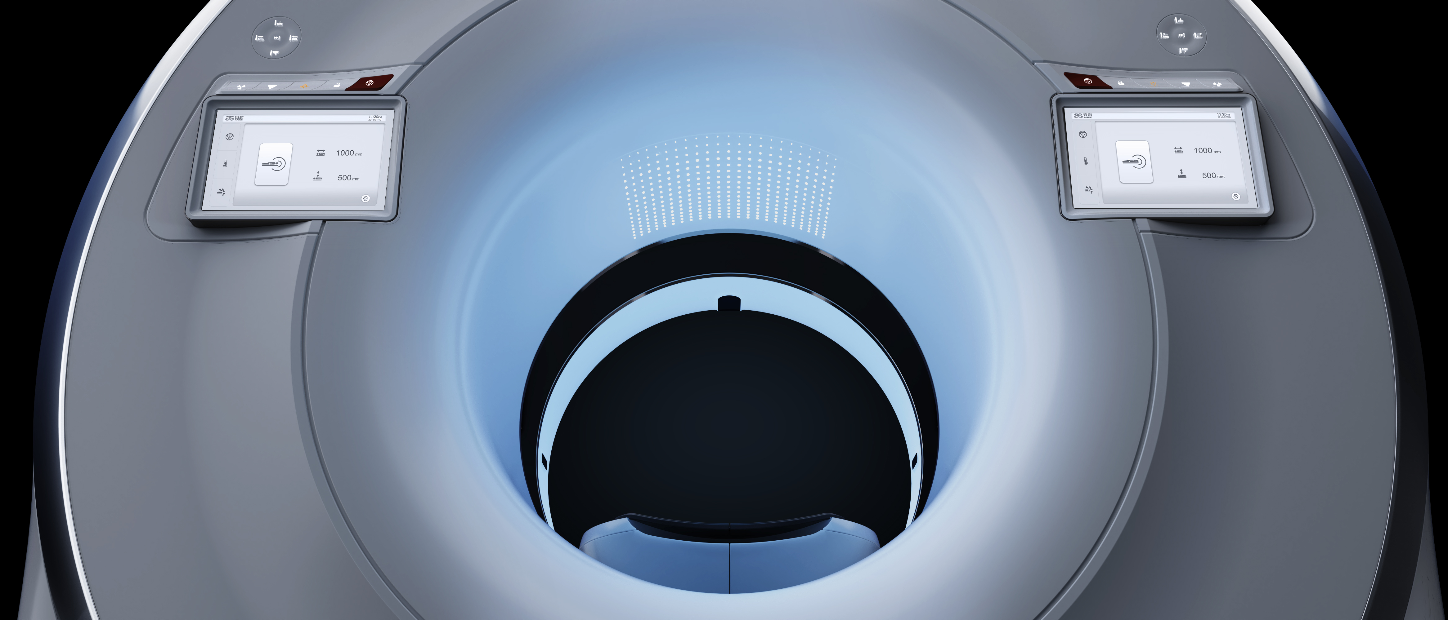
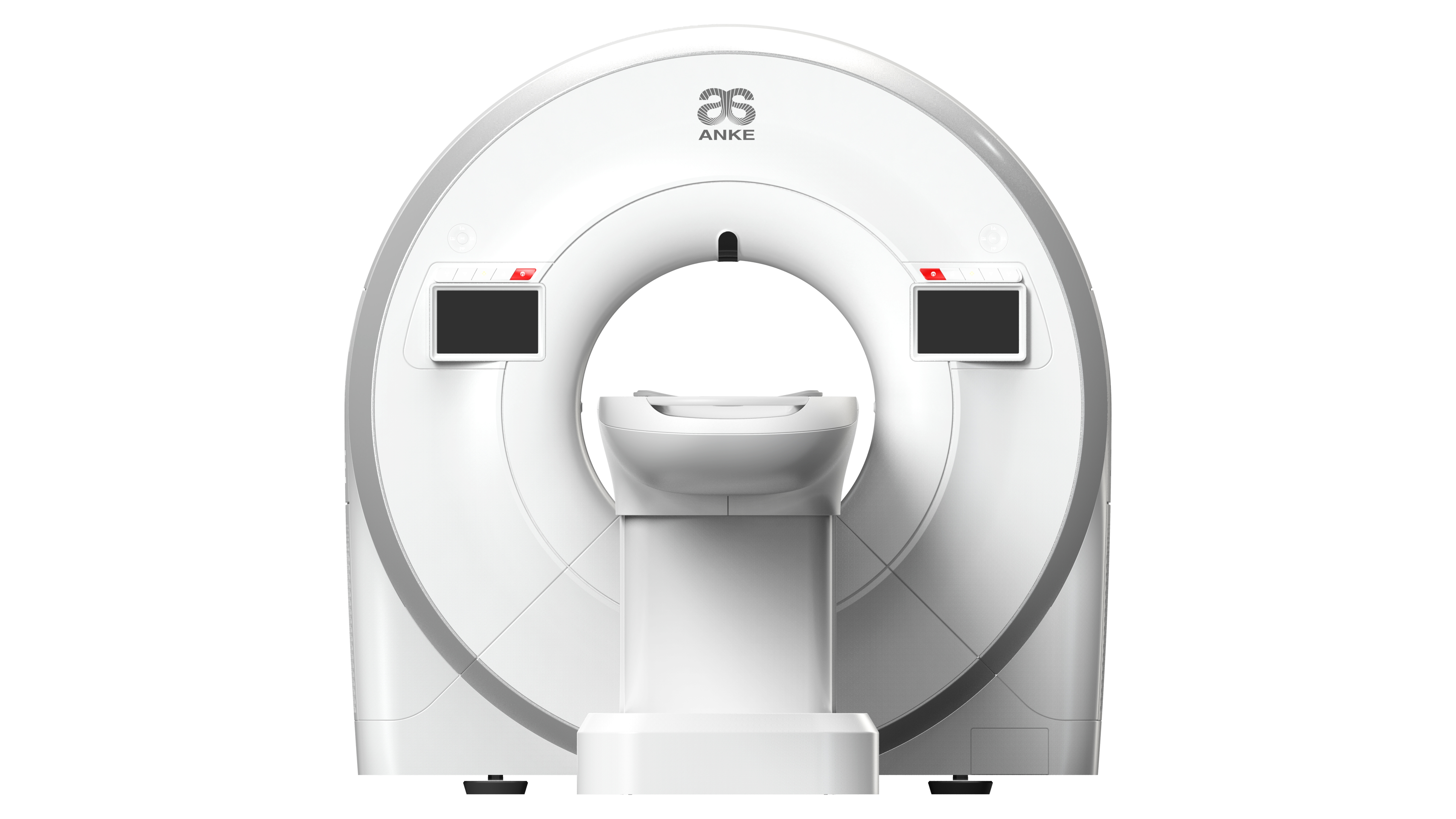
The ANATOM S400 with 76cm gantry aperture, combined with the unique design of the mood-soothing ambient lighting, minimises patient anxiety and nervousness, making it more conducive to performing procedures such as cardiac coronary examinations. In addition, the system offers four large, high-resolution touch screen
control panels, which not only provide the operator with a multi-view of the patient’s examination information and equipment status, but also allow for multi-modal patient positioning via control buttons and wireless remote control. It can minimize operator effort and improve examination efficiency, as well as enable contactless patient positioning.
Whith the help of a deep binocular visual perception system-Apps, deep learning is used to give the device the cognitive ability and behaviour to enable the system to intelligently identify multiple positioning points on the human body and display the scanning site on the intelligent touch screen terminal. As well it enables automatically identify the isocentric position of the proposed scanning site to achieve precise and intelligent patient positioning. Through the application of this technology, it not only substantially improves the accuracy of patient positioning and reduces operational errors, but also better protects patients from the risk of collision and accidental injury. More importantly, the Apps system also helps the technician to standardise operations and mitigate the increase in radiation dose to the patient’s surface due to inaccurate positioning, while further reducing image noise, reducing artefacts and improving image quality.
ANATOM S400 adopts ANKE’s self-developed 4-cm new generation OptiWave detector, which is organised and supported by the Ministry of Industry and Information Technology of the People’s Republic of China. It is designed with an isofocal hardware structure, combined with the AccuShape3D stereoscopic anti-scattering grids, so that the distance from the X-rays to the corresponding receiving unit of the detector is the same. In addition of obtaining high quality, high resolution images, it also achieves 4cm wide body coverage in both axial and spiral scanning, which not only improves examination efficiency but also enriches clinical applications.
The ANATOM S400’s fastest speed of 0.286 seconds/rotation, combined with the large-pitch HD fast scan technology, enables a scanning speed of 210 mm/s, which is more conducive to one-stop stroke examination, one-stop chest pain TRO examination, multi-site combined three-phase enhancement scan and whole-body oversized CTA examination, which not only effectively shortens the examination time and reduces radiation dose, but also significantly improves the success and image quality. It significantly improves the examination success rate and image quality as well. Additionally, the system offers the industry’s fastest reconstruction speed of 100 frames per second, providing doctors with a definitive diagnosis in the first instance and giving patients more time to save their lives.
With the advancement and development of CT technology, the use of CT diagnostic imaging is becoming more and more widespread, and the resulting radiation dose is of increasing concern to medical practitioners and the general public. How to achieve a reasonable radiation dose? How can the principle of dose minimisation be achieved? These are the main concerns of medical equipment manufacturers and imaging staff. Although iterative processing techniques have been introduced and used by all manufacturers, the enhanced image plasticity associated with high weighting iterations cannot be avoided. To better address this issue, the ANATOM S400 uses the Artist high-fidelity image noise reduction algorithm based on deep learning technology to optimise and process low-dose images. Compared to other algorithms, Artist enables a more thorough separation of noise from the image signal and ensures that no image detail information is lost during processing, resulting in high resolution clinical images at low doses.
Early detection, diagnosis and treatment have been agreed upon for many years, but achieving this is challenging, especially for small lesions (<0.6mm diameter), which are often missed due to the volumetric effect of thicker films. Although sub-millimetre acquisition and reconstruction is now possible with the vast majority of devices, the diagnosis is hampered by the low signal-to-noise ratio of thin-slice images. Based on the above situation, ANKE proposes the AI-based high-resolution thin-slice image reconstruction technology ASR (AI Super Resolution reconstruction), which uses a neural network processing unit to correct and reconstruct the artifacts of the overlapping projection data with artifacts by means of deep learning, which can significantly reduce the overlapping artifacts, and improve the resolution of CT images. By obtaining super thin-slice images with a high resolution of 0.3125mm, you will improve the accuracy of clinical diagnosis.
Spectrum imaging was implemented in 1973 by Hounsfield, the inventor of CT, by using two tube voltages for sequential scanning to distinguish between substances of different atomic number. Alvarez and Macovski, the first to explain the principle of CT dual energy imaging, pointed out that X-rays with mixed energy penetrate the body at the same time and that the Photoelectric and Compton effects produced in different substances can be used for CT Spectrum imaging.
There are two commonly used methods of X-ray energy decomposition, one is based on projection data domain, the other is image data domain. Studies have shown that the former is more accurate and facilitates the removal of artefacts, especially if the data is perfectly matched in time and space.
Currently, there are a number of proven CT energy imaging modalities available on market, including dual-source energy, high & low-kV switching, dual-layer detector and dual-helix/dual-axis.
Among them, dual helix/dual axis energy imaging has certain limitations (limited to sites or organs with autonomous motion) and is often used in low-end CT products; while dual source CT, dual layer detector CT and kV switching energy Spectrum CT can obtain good energy separation effect, but due to the high acquisition and maintenance costs, they cannot be widely used and promoted. In view of this, we have introduced the “Flexible Spectrum Imaging” technology based on conventional X-ray generation systems and OptiWave detectors.
The technology uses a flexible voltage switching scanning method to acquire data and uses AI-accelerated iterative reconstruction algorithms and energy reconstruction techniques to obtain energy spectrum images.
CCTA imaging, as the most important clinical application of post 64-row CT, has always been the most important technical point of interest for users during the procurement and use process. How can the success rate be improved? How can we make the examination more comfortable? How can we meet the needs of patients with high heart rates? How to meet the needs of patients with irregular heart rates? How to achieve low radiation dose and low contrast dosage for coronary imaging? Can energy spectrum imaging be applied to CCTA imaging? How can coronary artery analysis and diagnosis be performed quickly? How can we provide more accurate information to clinicians? …All these questions are the difficulties and pain points encountered by the majority of medical technicians in actual clinical work. In order to effectively solve these objective and practical problems, ANKE uses advanced hardware, new algorithms and leading AI imaging technology in the ANATOM S400 CT, taking advantage of the 8cm wide body detector, to bring patients a high success rate and high comfort of the examination experience, providing operators and clinicians with an easy, fast and comfortable way to diagnose and diagnose. It provides easy, fast and comfortable operation and diagnosis for both the operator and the clinician.
- AccuPitch coronary adaptive pitch technology, combined with AccuGating intelligent gating trigger technology and Adose mA current modulation technology, enables CCTA imaging at ultra-low dose and low contrast agent dosage
- Coronary artery examination supported by intelligent phase selection, intelligent premature beat skipping, intelligent ECG editing
- Perfectly adapted to patients with high heart rates and complex rhythms with the support of motion correction technology
- One-click coronary extraction and analysis, IVUS simulated intravascular ultrasound, plaque analysis, cardiac function analysis, calcification score analysis, vascular Endoscopy, CT-FFR technology for comfortable image processing and accurate diagnosis.
Typical Clinical Solutions
- Stroke solutions: including one-stop stroke screening, one-click brain haemorrhage analysis, ultra-low mA acquisition and Artist Intelligent image processing technology to ensure accurate data at low dose.
- Chest pain centre solution: covers the entire chest in 5-6 seconds, with intelligent ECG gating, intelligent mA technology and intelligent collimation adjustment to fully ensure scanning at low dose.
- Low-dose lung screening solution: Low-dose spiral CT has become the most effective screening tool for lung cancer. ANKE has achieved low-dose (ultra-low-dose) lung screening using Adose mA, Adose kV technology and the advanced Artist algorithm, combined with superior ASR high-resolution reconstruction technology and lung nodule analysis to ensure that small lesions are not missed and that doctors are more efficient in their diagnosis.
- Comprehensive lung analysis solution: In addition to the widely used lung nodule analysis function, ANATOM S400 CT also provides lung analysis functions such as high simulation bronchoscopy and intelligent pneumonia analysis to fully meet the clinical applications of respiratory departments and other departments.
- The Orthopaedics Direct solution: includes one-touch flesh and bone separation technology, internal bone fixation fluoroscopy, iterative and energy de-metallisation artefacts, spine and rib analysis technology and more. The spine analysis technology uses a deep learning approach, supported by big data, to automate spine extraction, identification and labelling, and to automatically reconstruct the optimal cut, thus freeing the doctor from tedious basic tasks in order to spend more time on complex cases.
